MODULE 3. Further laboratory and imaging examinations related to breast cancer diagnostics
3. Magnetic Resonance Imaging – MRI

This section covers imaging the breast with magnetic resonance imaging (MRI). After studying the material you will know what MRI is, and how it differs from other modalities. You’ll know the indications for breast MRI and when breast MRI is being used. You’ll know what the radiographer’s role is in MRI and how the radiographer guides the patient and performs the imaging process safely. You will find out how the patient is positioned for the examination and how the radiographer monitors the patient during the examination. You’ll know some of the techniques used in breast MRI and how the image is produced in MRI. The module also covers information about MRI-guided breast biopsy and contrast media used in MRI.
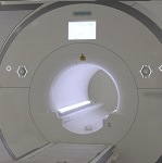
Hello, and welcome to this module about breast MRI. Magnetic resonance imaging of the breast, or breast MRI, uses strong magnetic fields to produce images of the internal structures of the breast and surrounding tissues. Breast MRI is used as a further examination method in addition to mammography.
In this learning module, we cover
The goal of this material is to improve cooperation between different professional groups, and ultimately, to serve better the women who enter the breast cancer diagnostic process.
We hope this material gives you tools to develop your skills, to understand what your fellow professionals are doing at the different stages of the diagnostic process, and to give the best possible care to your patients.
In this learning module, we cover
- The indications for breast MRI – when is a breast MRI used
- Technological principles and the variety of techniques – how does breast MRI work
- Image processing
- Positioning and patient guidance – how to get the best possible images
- Contrast agents
- Safety issues – including contraindications
- MRI guided biopsy
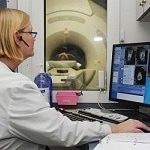
- And radiographers’ roles in breast MRI
The goal of this material is to improve cooperation between different professional groups, and ultimately, to serve better the women who enter the breast cancer diagnostic process.
We hope this material gives you tools to develop your skills, to understand what your fellow professionals are doing at the different stages of the diagnostic process, and to give the best possible care to your patients.