MODULE 3. Further laboratory and imaging examinations related to breast cancer diagnostics
3.7. MRI - guided breast biopsy
|
MRI-guided breast biopsy is a non-surgical medical procedure in which a tissue sample of the breast is taken under the guidance of MRI. This procedure is used when other imaging method cannot view breast lesions. Examining the tissue sample can help to differentiate between benign and malignant lesions. There are no absolute contraindications for MRI-guided breast biopsy, but it can be complicated by
|
|
Other common contraindications are the same as with any other MRI examination, or when using contrast media, or performing a biopsy:
- pacemaker
- claustrophobia
- coagulopathy
- allergy to local anaesthetic
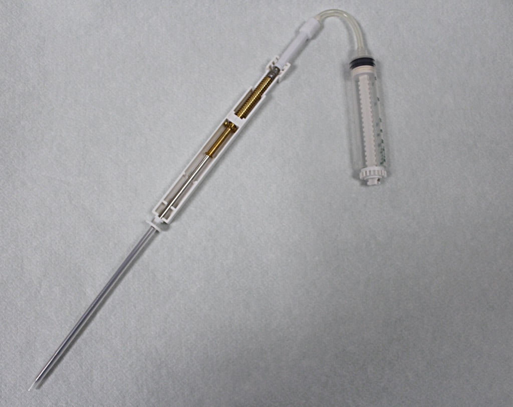
Breast biopsy
The biopsy technique depends on what kind of a sample is needed. Histological samples can be taken by core needle biopsy and cytological samples by fine needle aspiration. The most common technique is vacuum-assisted breast biopsy with an 11G needle. These techniques might involve taking several samples, the number of samples varying from 1 to 12.
The equipment must be of non-ferromagnetic material to avoid safety risks and artefacts.
First, an image is taken without the contrast agent to ensure fat saturation of the breast before injecting the contrast agent. For biopsies, a commonly used sequence is T1 weighted gradient echo sequence because it is relatively fast and therefore resulting in fewer artefacts due to motion. During biopsies, diagnostic images can also be taken.
The needle insertion site is swabbed with antiseptic and a grid is placed to help in finding the right insertion site for the needle. After the needle is in place, a control image is taken to ensure its correct location. The biopsy sample is taken by using vacuum and a twist motion.
The operation should be completed within 30 minutes after the contrast agent injection, as the quality of the sample decreases the longer the operation takes and the more the patient moves.
After the collection of a sufficient number of samples, the patient should lie on her back for fifteen minutes with a compression over the biopsy site(Plantade & Thomassin-Naggara 2014; Rissanen & Dean 2016).
Handling and processing of biopsy specimens
Specimens are handled according to the laboratory guidelines.
In a histopathological laboratory, the tissue samples are cut and transferred to tissue cassettes, fixed and sectioned, transferred to glass slides and stained, and examined. The pathologist writes a report about the examined breast lesion. The whole process takes around 3-5 days (Mäkinen 2012).
In a histopathological laboratory, the tissue samples are cut and transferred to tissue cassettes, fixed and sectioned, transferred to glass slides and stained, and examined. The pathologist writes a report about the examined breast lesion. The whole process takes around 3-5 days (Mäkinen 2012).
<PREVIOUS NEXT>
References
- Hukkinen K. Milloin rintojen MRI ja UÄ? Sädeturvapäivät abstraktit, 2012 (http://www.sadeturvapaivat.fi/index.php?id=688&cat_ids=x86x#cat86)
- Mäkinen M. Näytteiden käsittely laboratoriossa. Patologia toim. Duodecim, 2012.
- Plantede R, Thomassin-Naggara I. MRI vacuum-assisted breast biopsies. Diagnostic and Interventional Imaging. 2014; 95: 779-801.
- Rissanen T, Dean PB. Ohjauksessa otetut neulanäytteet. Duodecim, 2016 (http://www.oppiportti.fi/op/krd00906/do#s4)
