MODULE 3. Further laboratory and imaging examinations related to breast cancer diagnostics
3.4. Positioning and patient guidance – how to get the best possible images
Positioning the patient
|
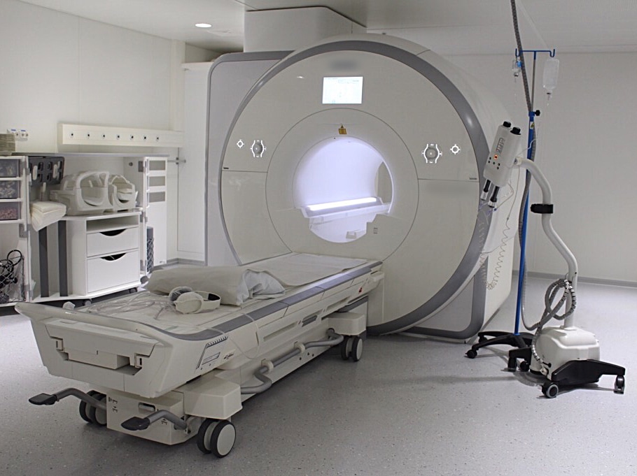
After preparing the patient for the examination, the patient is led to the examination room.
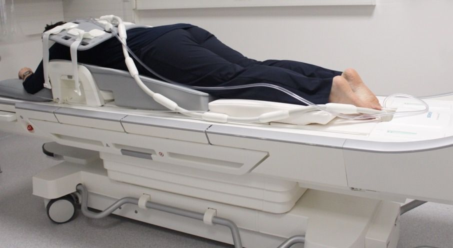
The patient is guided to take a prone position on the examination table so that her breast is hanging through the hole in the breast coil, with her arms over the head (Price & Morris 2011). The correct position of the breast inside the breast coil has to be checked before starting the examination. The breast should be positioned in the middle of the coil. Both lateral and medial directions should be checked. To prevent any artefacts in the images, the nipples should be positioned in profile (Price & Morris 2011). It is important to make sure that the patient is comfortable before starting the examination, because the examination usually takes approximately a half an hour and the patient is not allowed to move at all during that time. |
|
Reasons for an unsuccessful examination can include
- Patient movements during the examination
- Insufficient positioning of the breast
Before the start of examination, the patient gets headphones to minimize the noise and have an the opportunity to listen to music during the examination. For emergencies, the patient is given a call button to allow the patient to contact the radiographers if she experiences any problems during the examination (Price & Morris 2011).
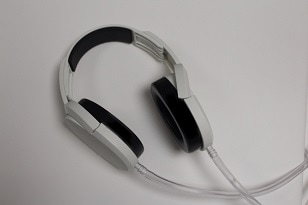
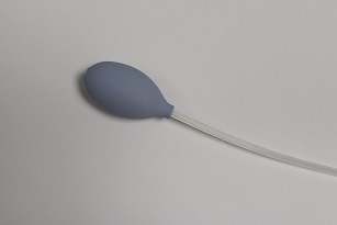
MRI scanning is very loud, so patients should wear hearing protection devices during the scan. The patient can call the radiographer using the call button, if needed (Polus 2012; Tallqvist et al 2015).
During the examination
|
During the examination, the radiographer monitors the patient from the next room through a window.
The radiographer is ready to interrupt the examination if the situation demands it. The radiographer talks to the patient and tells her about the duration of the examination and motivates the patient to remain still to ensure the diagnostic value of the images. The examination is discontinued in case the patient is too restless to continue and the patient receives a new examination time. If needed, the patient receives instructions on how to proceed at home after the examination. These instructions may inform on how to take care of themselves after a biopsy, the administration of contrast agent or other medication (Polus 2012). |
Note that watches are not allowed in the MRI room. The photo above is an example.
|
<PREVIOUS NEXT>
References
- Polus P. Potilasohjekansio magneettitutkimukseen tulevalle aikuispotilaalle. 2012.
- Price, Elissa R. – Morris, Elizabeth A. Magnetic resonance imaging- guided breast biopsies: tips and tricks. Canadian Association of Radiologists Journal. 2011; 62 (1): 15–21
- Tallqvist – Tolonen - Ylifrantti. Potilasturvallisuus magneettikuvantamisessa. Radiologia. 2005; Rintojen MRI 5.2.7: 250
